Chordoma is a rare tumour which is thought to arise from remnants of the embryonic notochord (cartilage). Generally, it is slow-growing, although rarely, some chordomas can be more aggressive. It can grow anywhere along the spine column. Today we will discuss skull base chordomas specifically due to the complex treatment strategy required.
Managing base of skull chordoma typically involves a multi-disciplinary approach, combining surgery and radiation therapy. The anatomy is challenging due to close-ness to critical structures like the cranial nerves, eyes and brain. Here are some key aspects of the management of base of skull chordoma:
- Surgery:
- Maximal Safe Resection: The primary treatment for chordomas is surgical removal. However, complete resection can be challenging due to the tumor’s proximity to critical structures, such as the brainstem, nerves, and blood vessels.
- Skull Base Approaches: Surgeons often use specialized skull base surgical techniques to access and remove the tumor without causing excessive damage to surrounding structures.
- Radiation Therapy:
- Adjuvant Radiation: Even after surgery, residues of the tumor may remain. Adjuvant radiation therapy is suggested to target tumor cells left behind.
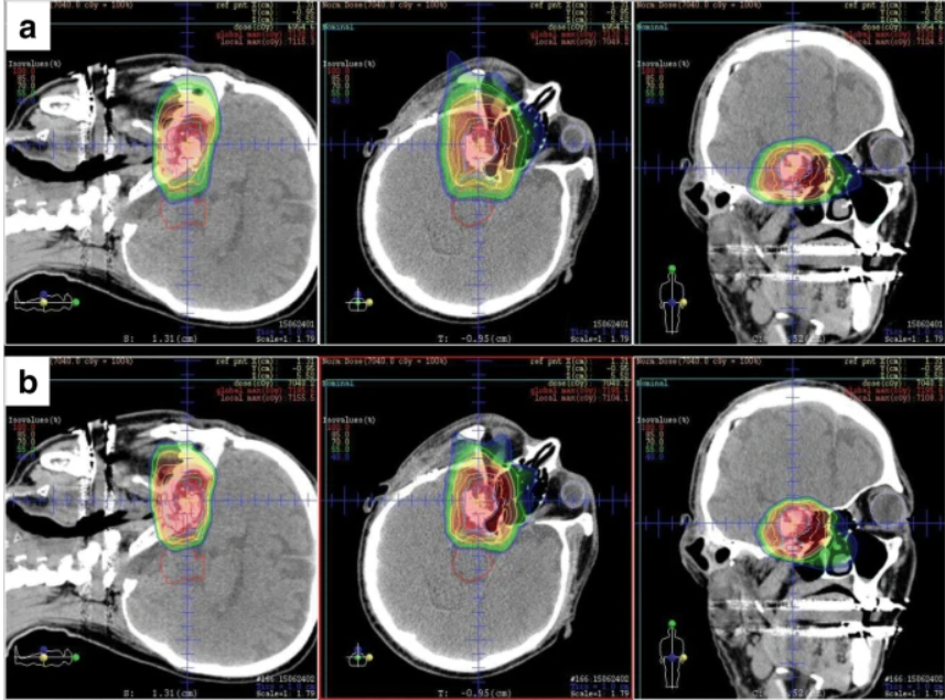
- Proton Beam Therapy: Due to the location of base of skull chordomas, proton beam therapy may be preferred. Proton therapy delivers radiation more precisely, minimizing damage to surrounding healthy tissues.
Modern radiotherapy techniques, particularly with carbon or proton beam irradiation, have yielded excellent local control rates, even in the presence of large disease due to surgical challenges. There have been studies published by Japanese groups comparing carbon and proton beam. Outcomes are similar.
In this Japanese study published in 2018, both patients who had and did not have surgery were studied after treatment with carbon or proton beam therapy. At 5 years, local control was 85%, and results suggest patients who had good removal of tumour had better outcomes. In a more recent study published by doctors from the University of Florida using a more precise proton technology (pencil beam) reported that local control was 95% when patients had total removal versus 70%. There were no serious toxicities due to radiotherapy.
To coordinate efforts to manage this challenging and rare disease, large international groups have collaborated to publish guidelines for doctors. This includes the Global Chordoma Consensus and the ESTRO radiotherapy treatment guide.
We recommend engaging the neurosurgeons and radiologists, together with radiotherapy physicists to develop a robust and safe radiotherapy plan.